NewsReseach Result
Approach from a new perspective: Raman microscopy visualizes hepatic molecules in living cells under drug administration.
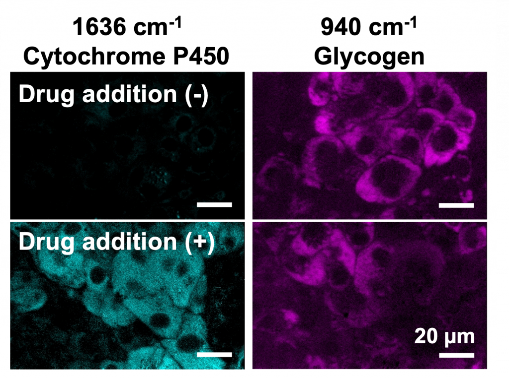
Using a line-illumination Raman microscope, which can observe a sample several hundred times faster than conventional techniques, we have successfully observed the effects of drugs on cytochrome P450 (CYP) activity and glycogen accumulation in a non-destructive and label-free manner. This achievement will lead to a more accurate interpretation of drug effects by evaluating cellular responses under natural conditions. This technique is expected to be useful for new drug development and quality control of cellular products in regenerative medicine.
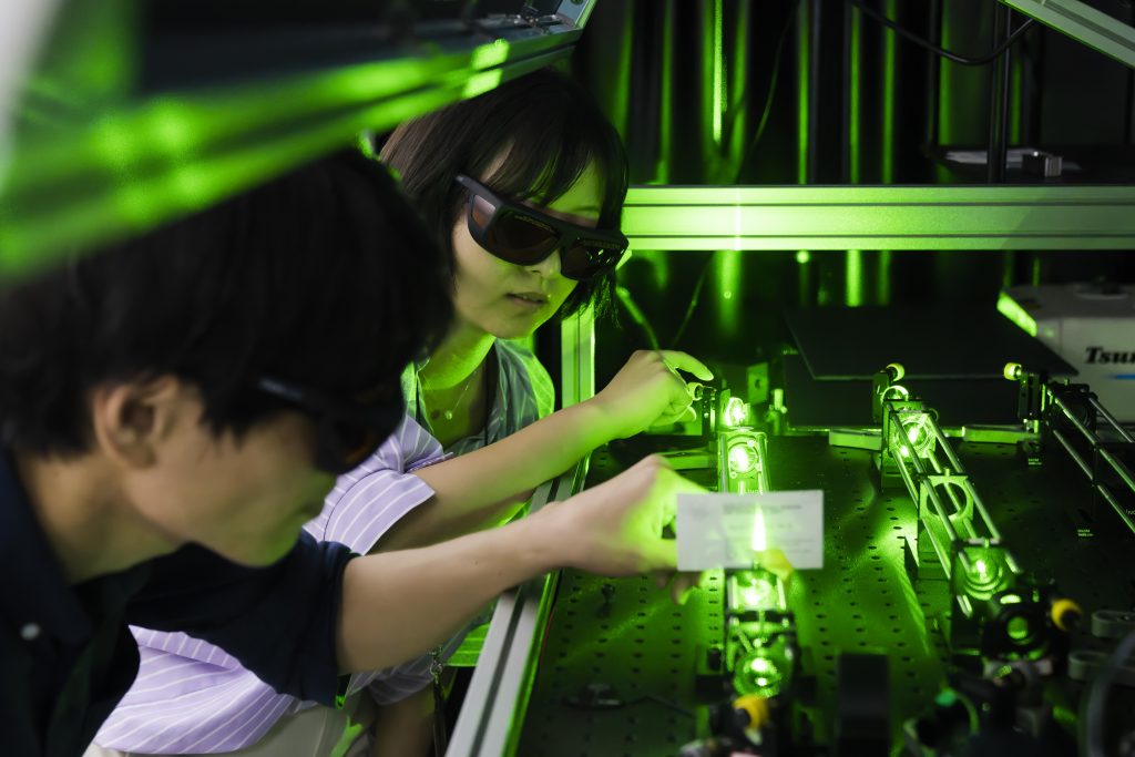
Fujita Laboratory in Department of Applied Physics is developing a high-speed Raman microscope applicable to live cell imaging. The first author, Dr. Menglu Li, a specially-appointed assistant professor in Department of Applied Physics (obtained her Ph.D. from Department of Biotechnology, Graduate School of Engineering, Osaka University), found the Raman spectroscopic imaging technique useful from a biological perspective and, together with co-first author Dr. Yasunori Nawa (now at Kwansei Gakuin University), successfully observed the activity of CYP without modifying cell conditions. This is the first successful label-free observation of CYP activity. This research was also performed in Advanced Photonics and Biosensing Open Innovation Laboratory, which was established to integrate bioanalysis and photonics technologies.
Raman microscopy is expected to be a useful tool for research in the fields of biology and medicine because it allows the observation of biomolecules with analyzing them. However, its potential is still unknown, and it is not rare to obtain surprising results from biological samples that are observed for the first time. Although the technology is still in its developmental stages, Raman microscopy is a fascinating tool that allows us to view living organisms from new perspectives and to make new discoveries in biology.